01Concept design · Laparoscopic
Cryogel adhesion removal
Freeze a post-surgical adhesion band in a dye-loaded gel, then crack it with near-infrared laser pulses instead of cutting it.
Coursework concept · Not built or tested01 · Coat. A laparoscopic instrument sprays ICG-loaded alginate cryogel onto the adhesion band between intestine and abdominal wall.
The concept sequence, drawn from the course report. Schematic, not to scale.
- Course
- BME 250, University of Illinois Chicago
- Timeline
- Fall 2025 · about 5 weeks
- Team
- 4 students
- My part
- Material, structure and laser-parameter selection; safety review
01Problem
Adhesions form in over 90% of patients after intra-abdominal surgery. These bands of scar tissue tie the intestine to itself, to other organs or to the abdominal wall, and can cause pain and bowel obstruction. Cutting them takes another operation, and they can grow back. We set out to break bands apart without cutting them.
02Requirements and constraints
Delivered laparoscopically through a standard 5 mm port, for bands up to Zühlke grade 3. Grade 4, where organs are fused, was excluded for safety. Neighboring organs must not freeze or burn. Four requirements, each with a test, are in the table below.
| Requirement | Planned test | Status |
|---|---|---|
| Cryogel is biocompatible | Cytotoxicity test | Not run |
| Cryogel freezes to −18 °C | Temperature measured with a thermometer | Not run |
| Cryogel absorbs the laser’s 800 nm light | Spectroscopy, Beer–Lambert law | Not run |
| Laser breaks adhesions | Procedure on scar-tissue models of varied thickness and density | Not run |
03Design decisions and why
- 01Match the light to a dye, not to tissue.
- Indocyanine green (ICG) absorbs near 800 nm, the output of common diode lasers, and water-rich tissue absorbs little there. Loading the gel with ICG puts the energy in the gel (selective photothermolysis).
- 02Make the gel brittle on purpose.
- EDC crosslinking forms short, rigid “zero-length” bonds, and in our source data EDC/NHS gave the largest mean pore size of five crosslinkers (165 ± 86.5 µm). Rigid bonds and large pores favor fracture. Alginate is biocompatible and inexpensive.
- 03Crack by thermal shock, not ablation.
- Pulses of 0.1–10 ms heat the gel’s surface while its core stays below −5 °C, so the warm shell expands against the frozen core and the band fractures.
- 04Reuse surgical hardware.
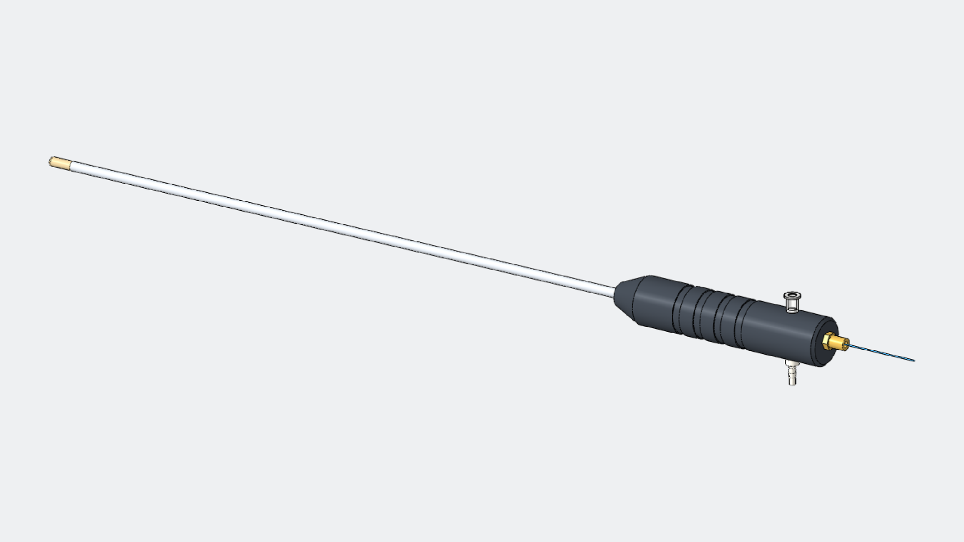
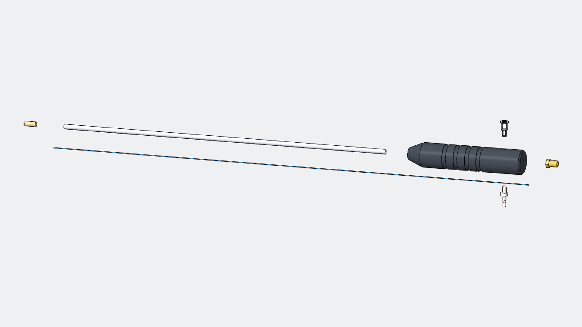
- The laser stays outside the body. A 0.2–1 mm fiber runs through the port in a three-lumen instrument that sprays gel, delivers light and suctions fragments.
04What I did
I selected the materials, device structure and laser parameters from the primary literature, and reviewed usability and safety during surgery, including the risk of freezing or burning nearby organs. I also modeled the three-lumen instrument in SOLIDWORKS 2025 (see CAD).
05Outcome and limits
A concept report; nothing was built or tested. On review, three gaps stand out: nothing names what cools the band to −18 °C inside a 37 °C abdomen, no laser power or pulse energy is given, and the pore-size data comes from chitosan/collagen hydrogels, not alginate cryogels.
06What I’d do next
- Add a cooling source: an argon Joule–Thomson cryoprobe (1.5–3 mm shafts, already used in cryosurgery) through a second port.
- Measure the gel’s absorbance, then set pulse energy on gel phantoms before any tissue.
07CAD
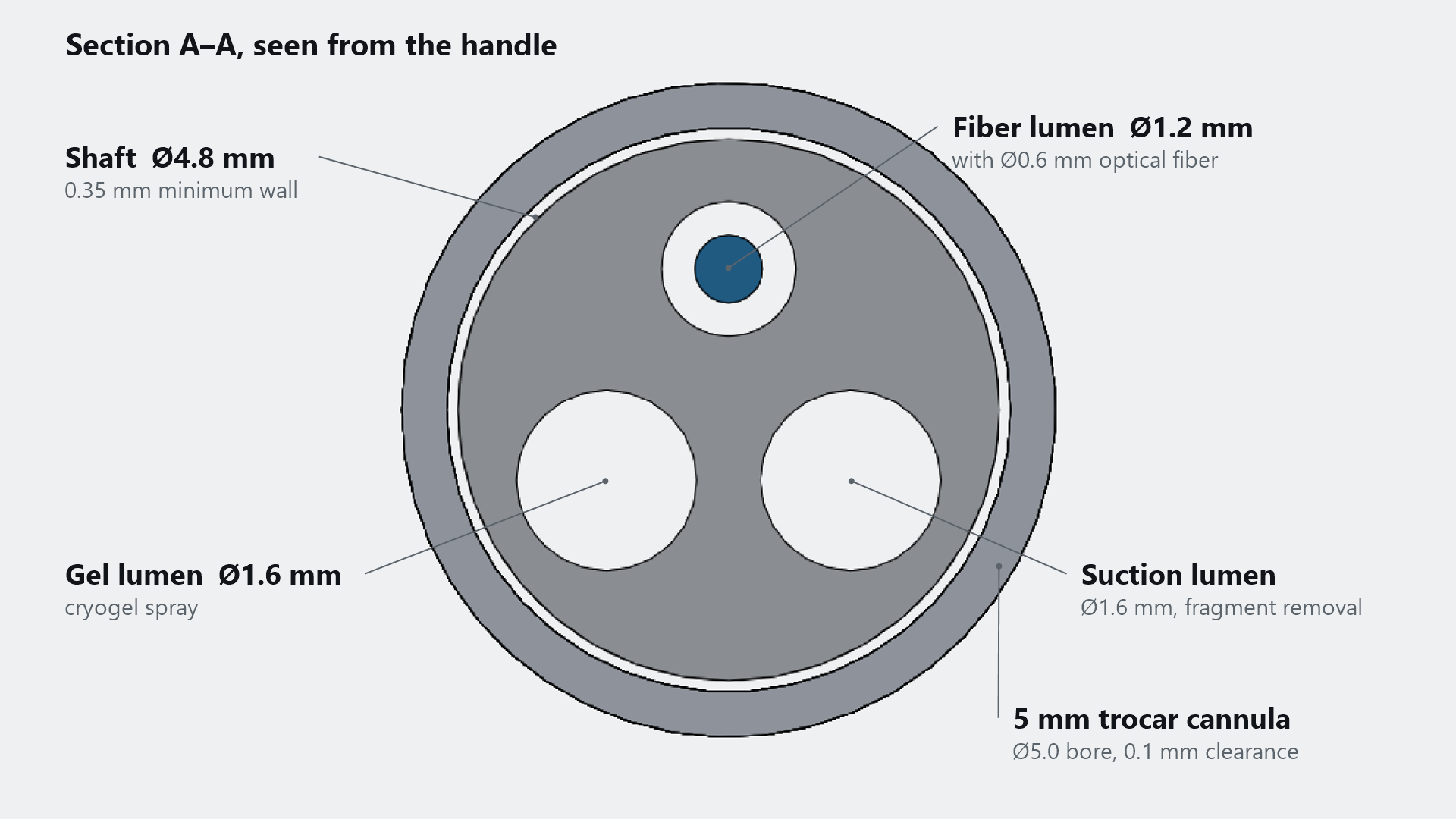
My SOLIDWORKS 2025 model of the three-lumen instrument: distal tip, shaft, handle, gel, fiber and suction ports, and the optical fiber. Section A–A checks the fit. A 4.8 mm shaft holds all three lumens with walls of at least 0.35 mm, and clears a 5 mm trocar by 0.1 mm on each side. Where the coursework gave no dimension, the value is an assumption.
Sources
Show all 7 sources
Cited in the course report
- Stommel MWJ et al. Efficacy and safety of the C-Qur Film Adhesion Barrier (CLIPEUS trial): study protocol. Trials 15, 378 (2014). doi:10.1186/1745-6215-15-378
- Sánchez-Cid P et al. Effect of different crosslinking agents on hybrid chitosan/collagen hydrogels. Int J Biol Macromol 263, 129858 (2024). doi:10.1016/j.ijbiomac.2024.129858
- Henderson TMA et al. Cryogels for biomedical applications. J Mater Chem B 1, 2682–2695 (2013). doi:10.1039/C3TB20280A
- Lee KY, Mooney DJ. Alginate: properties and biomedical applications. Prog Polym Sci 37, 106–126 (2012). doi:10.1016/j.progpolymsci.2011.06.003
- Pradhan S et al. Fundamentals of laser-based hydrogel degradation. Adv Healthc Mater 6, 1700681 (2017). doi:10.1002/adhm.201700681
- Gong Y, Li Y, He S. Thermo-mechanical weathering in Malan loess under thermal shocks. Sensors 25, 3115 (2025). doi:10.3390/s25103115
Post-project review
- Theodorescu D. Cancer cryotherapy: evolution and biology (argon Joule–Thomson probes, 1.5 mm and 3 mm). Rev Urol 6(Suppl 4), S9–S19 (2004). PMC1472868