02Concept design · Drug delivery
AI-guided tenecteplase auto-injector
An at-home IV injector for clot-busting medication that maps the vein with an RGB-NIR camera before it fires.
Coursework concept · Not built or tested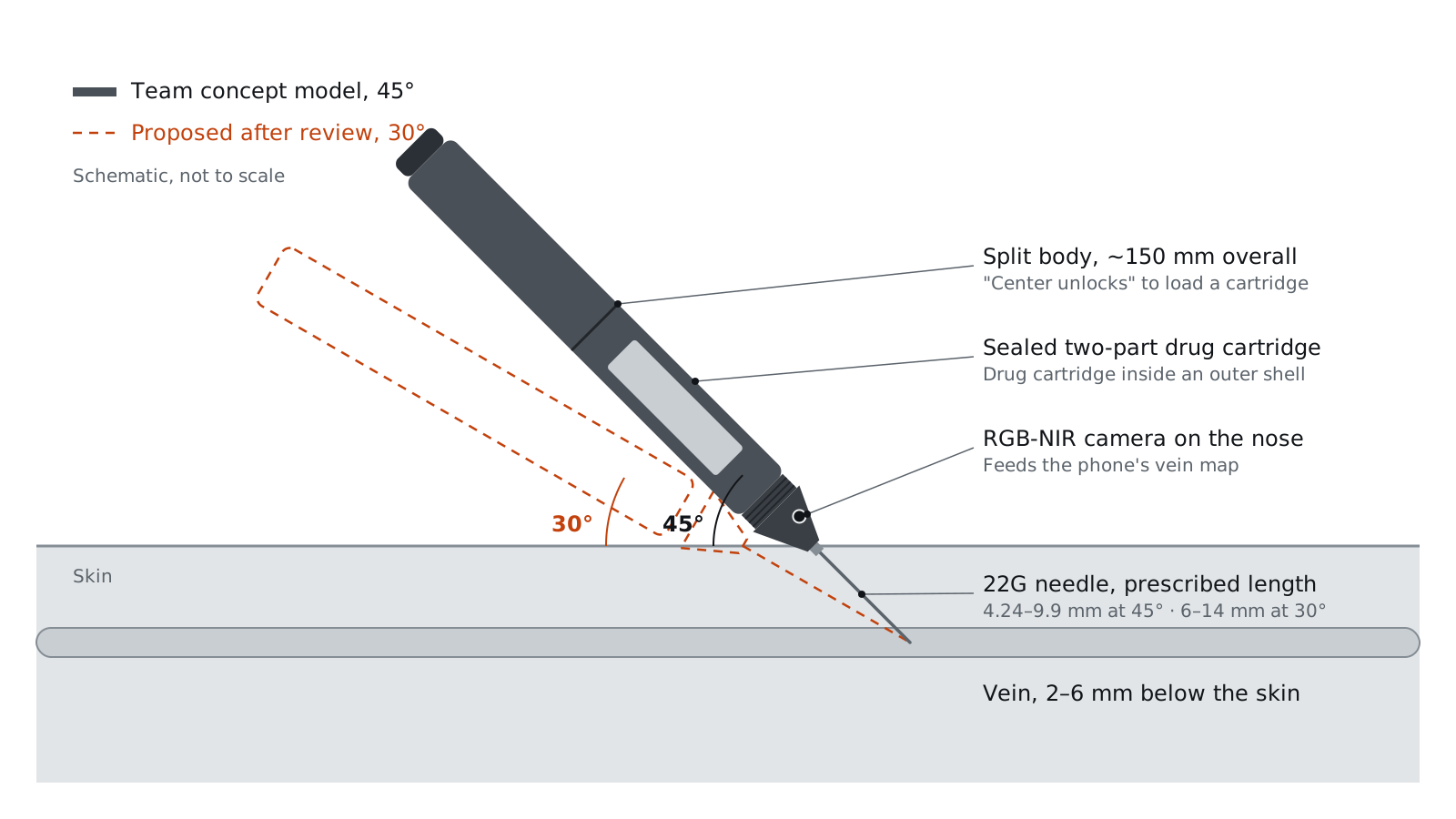
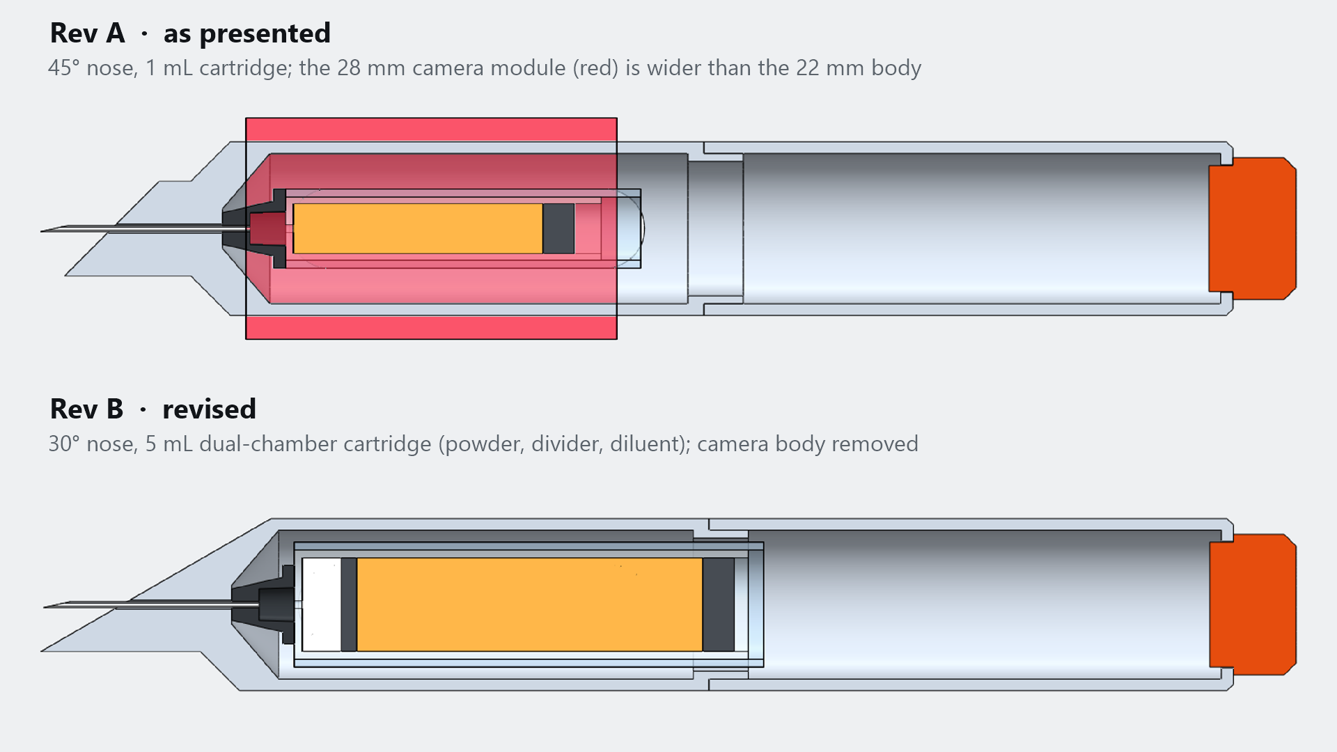
Layout from the team's concept model, with the 30° revision proposed after review. Schematic, not to scale.
- Course
- BME 250, University of Illinois Chicago
- Timeline
- Fall 2025 · about 5 weeks
- Team
- 4 students
- My part
- Vision-guidance subsystem, risk assessment, user-workflow map
01Problem
Clot-busting drugs work best within about three hours of symptoms, but the common ones run as IV infusions lasting hours, and at-risk patients often cannot reach a hospital in time. The brief: a clot-busting system an at-risk person can keep at home.
02Requirements and constraints
One rapid IV dose of tenecteplase, with dose and needle length prescribed by the patient’s physician; usable without phlebotomy training; a sterile single-use cartridge and needle. Verifiable requirements are in the table.
| Requirement | Planned test | Status |
|---|---|---|
| Needle retracts automatically without excessive force | Press the injector; confirm the needle retracts on its own | Not run |
| Insertion angle preset at 30–45° (30° after review) | Place on a flat surface and measure the needle angle | Not run |
| The app locates the vein and shows it on the phone | Photograph the app’s output on an arm; check the vein location with ultrasound | Not run |
03Design decisions and why
- 01Inject, don’t inhale or implant.
- An EpiPen-style injector beat an implanted monitor, a public injection station and an inhaler on portability and cost.
- 02Size the needle to the patient.
- A 22G needle (0.7 mm) comes in five lengths, 4.24–9.9 mm: a 2–6 mm skin-to-vein depth, plus 1 mm to the vein’s center, divided by sin 45°.
- 03Let the device see the vein.
- Hemoglobin absorbs near-infrared light differently from tissue, so veins stand out. An RGB-NIR camera (450–800 nm bands) feeds VV-Net, a lightweight neural network already run on phones, and the app shows the vein map before the user fires.
- 04Keep the drug sealed.
- A sealed cartridge sits inside an outer shell; a Luer-lock needle swaps out after each use.
04What I did
I owned the vision subsystem: I chose the camera, based guidance on VV-Net, and proposed a micro-ultrasound probe and a needle-tip impedance check to confirm vein entry. I ran the risk assessment (injection accuracy, sterility, drug storage, user workflow), mapped the user’s steps to find use errors, and turned both into requirements. I also rebuilt the team’s concept in SOLIDWORKS 2025, as presented and as revised (see CAD).
05Outcome and limits
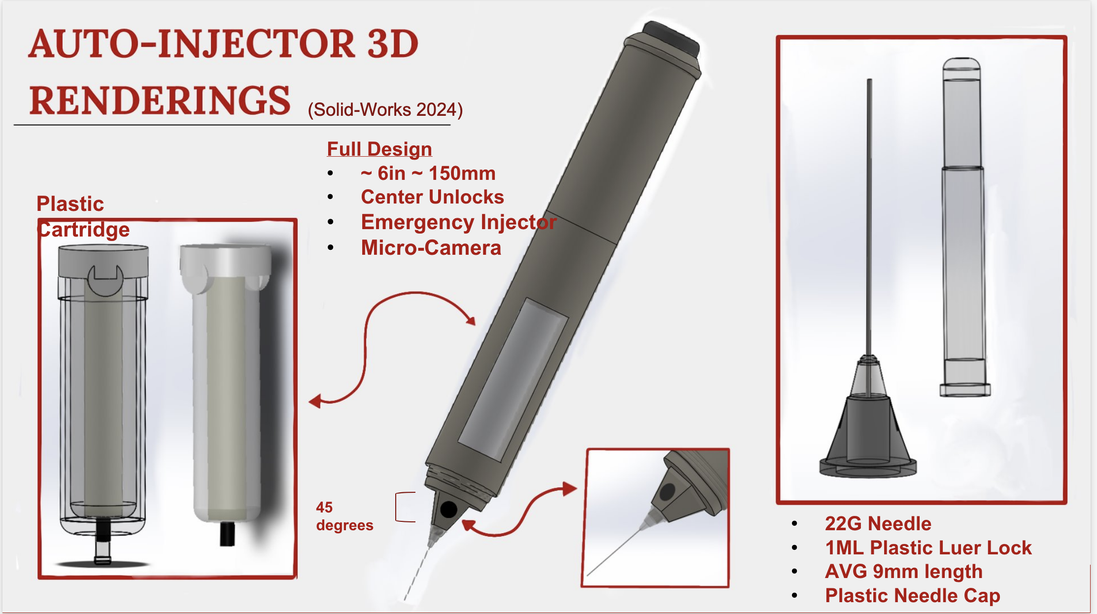
A concept report and a team-built SOLIDWORKS 2024 model; nothing was built or tested. On review, its numbers disagreed (angles of 15–30°, 30–45° and 45°; 1 mL against 5 mL), the render’s proportions put the pen near 23 mm across, narrower than the 28 mm camera module, and stroke thrombolysis normally follows a scan that rules out bleeding, which no home device can do.
06What I’d do next
- Fix the angle at 30°, the upper limit in IV-insertion guidance: a 2 mm vein gives a 4.0 mm window along the needle, against 2.8 mm at 45°.
- Size a dual-chamber cartridge to the label’s 5 mL maximum; the drug ships as powder and must be used within 8 hours of mixing.
- Redo the pressure estimate (about 8 psi, not 1.5) and replace the camera body with a bare sensor.
07Engineering checks
| Depth d | L at 45° (team) | L at 30° (proposed) |
|---|---|---|
| 2 mm | 4.24 mm | 6.00 mm |
| 3 mm | 5.66 mm | 8.00 mm |
| 4 mm | 7.07 mm | 10.00 mm |
| 5 mm | 8.49 mm | 12.00 mm |
| 6 mm | 9.90 mm | 14.00 mm |
| Window along the needle in a 2 mm vein, 2 ÷ sin θ | 2.83 mm | 4.00 mm |
Why the angle matters here: at the elbow, the median cubital vein sat 3.0 ± 1.8 mm deep and the brachial artery a median 3.6 mm beneath it in an ultrasound study, so a steep needle that passes through the vein’s back wall has little margin.
| Term | 9 mm needle | 14 mm needle |
|---|---|---|
| Viscous loss, Hagen–Poiseuille | 1.9 psi | 2.9 psi |
| Entrance loss (K = 0.5, assumed) | 2.1 psi | 2.1 psi |
| Exit loss, ½ρv² | 4.2 psi | 4.2 psi |
| Total | ≈ 8.1 psi (56 kPa) | ≈ 9.2 psi (63 kPa) |
Inputs: 1 mL/s, the label’s maximum 5 mL bolus over 5 seconds; 22G regular-wall inner diameter 0.41 mm; viscosity of water, 1 mPa·s (assumed). The jet leaves the tip at about 7.6 m/s (Reynolds number ≈ 3,100), so the losses at each end of the needle dominate and the total is an estimate. The report’s 1.5 psi matches the viscous term alone for a thinner-walled needle.
08CAD
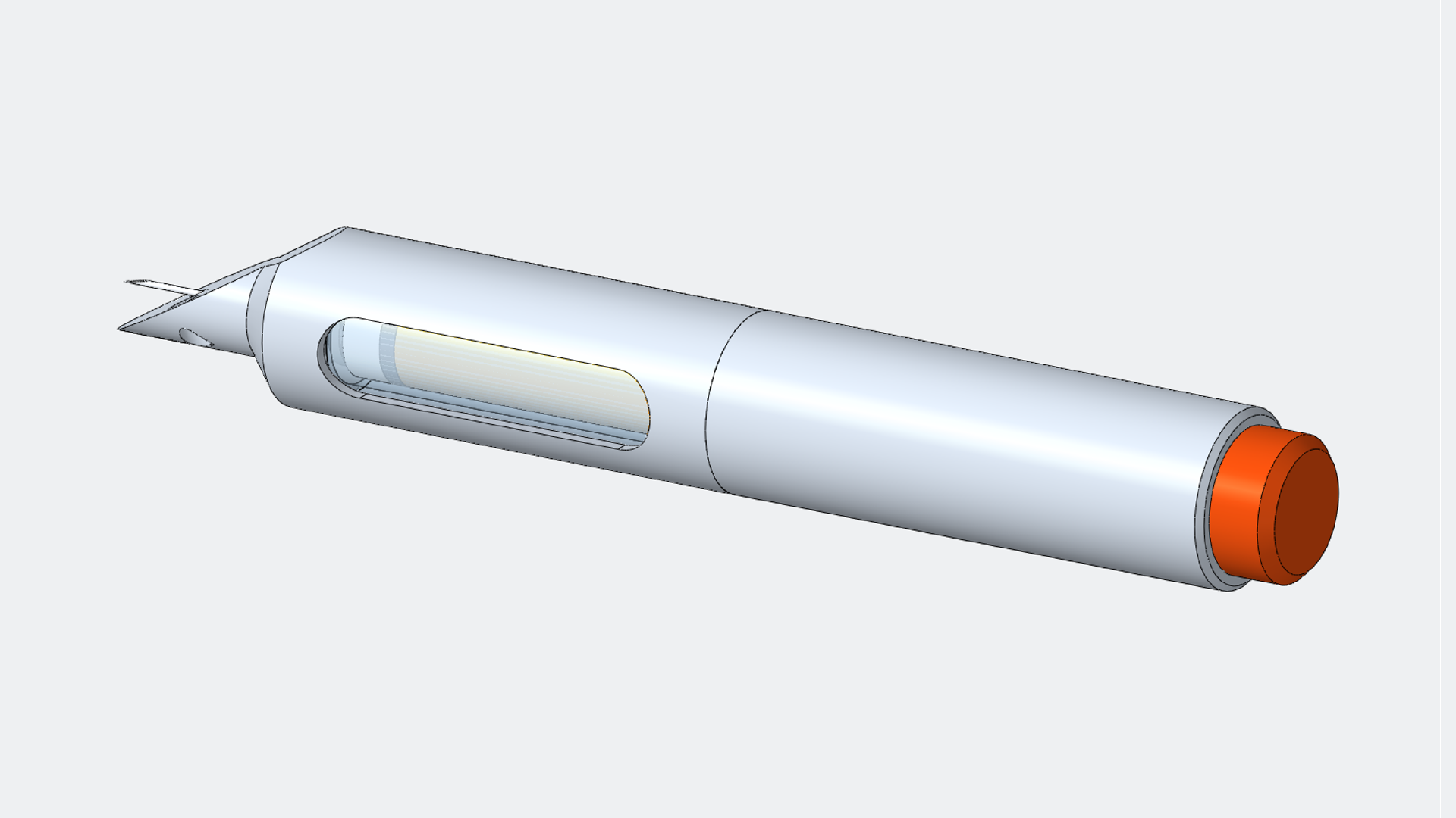
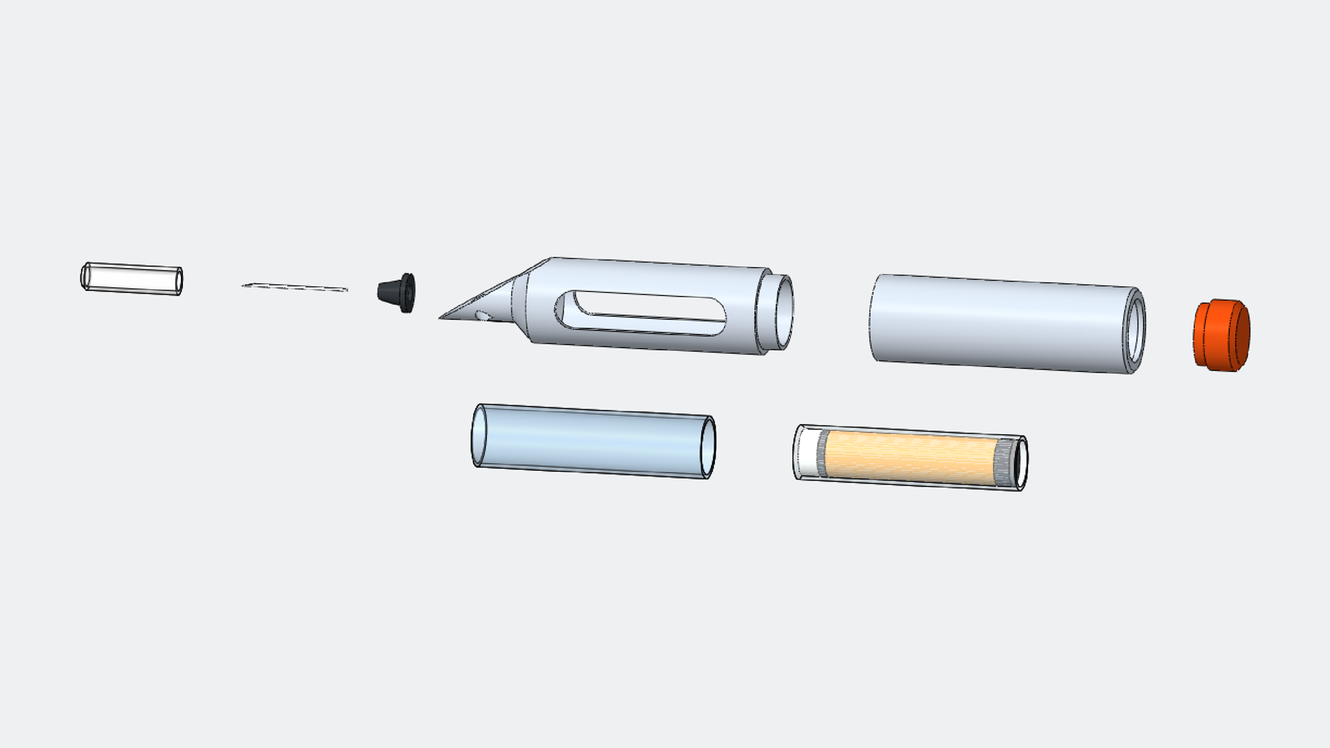
My SOLIDWORKS 2025 rebuild of the team’s concept: housing, nose, cartridge, needle hub, needle and cap, in two configurations. As presented: 45° nose, 1 mL cartridge and the 28 mm camera module. Revised: 30° nose, a 5 mL dual-chamber cartridge, and no camera body. Where the coursework gave no dimension, the value is an assumption.
Sources
Show all 10 sources
Cited in the course report
- Gill HS, Prausnitz MR. Does needle size matter? J Diabetes Sci Technol 1(5), 725–729 (2007). doi:10.1177/193229680700100517
- Mukai K et al. Safety of venipuncture sites at the cubital fossa as assessed by ultrasonography. J Patient Saf 16(1), 98–105 (2020). doi:10.1097/PTS.0000000000000441
- American Heart Association. New clot-busting medication simplified stroke treatment in specialized ambulance (2024). newsroom.heart.org
- Tang C et al. Deep learning-based vein visualization and its mobile deployment (2025). doi:10.1016/j.bspc.2024.107272
- Spectral Devices. RGB-NIR camera. spectraldevices.com
Post-project review
- Genentech. TNKase (tenecteplase) prescribing information: stroke dose 0.25 mg/kg up to 25 mg (5 mL), single bolus over 5 seconds; 5 mg/mL after reconstitution; use within 8 hours. gene.com
- MSD Manual Professional. How to insert a peripheral intravenous catheter (insertion at 10–30°). msdmanuals.com
- World Health Organization. WHO guidelines on drawing blood: best practices in phlebotomy (2010). who.int
- Powers WJ et al. Guidelines for the early management of acute ischemic stroke: 2019 update. Stroke 50, e344–e418 (2019). doi:10.1161/STR.0000000000000211
- ISO 9626:2016. Stainless steel needle tubing for the manufacture of medical devices (22G nominal sizes).